Question
Biology
What can a cyanobacterial micrograph reveal about prokaryotic structure?
Answer
Biology
Expert Answer
A cyanobacterial micrograph can show cell shape and boundaries. If an electron image has enough detail, it can also show how internal membranes are arranged. These features can help you identify a prokaryotic cell, but not every biological property can be read directly from the image.
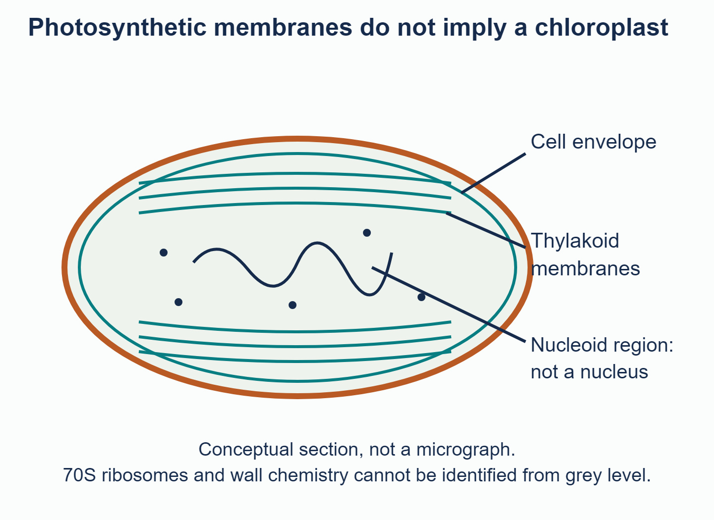
A cyanobacterium is prokaryotic even though it has internal photosynthetic membranes; the nucleoid is not membrane-enclosed.
Cyanobacteria are photosynthetic bacteria. Their DNA is not enclosed in a membrane-bounded nucleus. They can contain photosynthetic membranes, commonly arranged as thylakoids within the cell, but these are not enclosed inside chloroplasts. Photosynthesis therefore does not by itself identify a cell as a plant cell.
In a light micrograph, start with shape and size from the scale bar. Look for colonies or chains of cells, called filaments, and any visible colour or specialised cells. Ordinary light microscopy will not normally distinguish individual ribosomes, the plasma membrane's lipid bilayer (its two layers of lipids) or detailed DNA organisation. If you cannot see a nucleus, the image may lack detail or the section may miss it. That is weaker evidence than a clear view of how the cell is organised.
An electron micrograph can reveal the cell envelope, internal membrane outlines and a finely speckled interior. Even then, keep what you can see separate from evidence about chemical composition:
| Observation or claim | What supports it? |
|---|---|
| Repeated internal membrane profiles | Fine structural detail visible in a suitable section |
| No membrane-enclosed nuclear region in the section | Careful inspection, interpreted with the specimen identity |
| Wall contains peptidoglycan | Knowledge or analysis of bacterial wall chemistry, not grey level alone |
| Ribosomes are 70S | Laboratory analysis; "70S" is not a size directly read from pixels |
The diagram is an original explanatory schematic. Its nucleoid label represents the region containing genetic material; it does not imply that a routine micrograph reveals a neatly outlined DNA bundle. Bacterial DNA associates with proteins, so "naked DNA" should not be taken literally.
To make a strong answer, describe one visible feature and explain how independent biological knowledge supports its interpretation. Avoid a rigid size rule such as "anything below ten micrometres is a bacterium": sizes overlap and exceptions occur.
Finding a cell wall, photosynthetic membranes or chlorophyll does not mean you have found a chloroplast. Look at the overall organisation: cyanobacteria photosynthesise within a prokaryotic cell, not within a membrane-enclosed plastid inside a eukaryotic cell.
Explore the cyanobacterial image in the Biology Unity and Diversity questionbank. Choose features that support identifying it as a bacterium, making clear which you can see and which depend on your biological knowledge.
Answered by Revision Village IB Expert
Explore More IB Biology Resources
Over 80% of IB students globally are experiencing the power of Revision Village