Question
Biology
What can electron micrographs show that light micrographs cannot, and what do they still hide?
Answer
Biology
Expert Answer
Electron micrographs can resolve fine cellular structures that an ordinary light microscope cannot separate, such as the internal membranes of mitochondria and chloroplasts. Their advantage is resolution, not merely that the image can be printed larger.
Resolution is the ability to distinguish nearby features. Enlarging two merged spots produces a larger blur; it does not reveal that there are two. Conventional light microscopy typically resolves features separated by roughly under favourable conditions. Electron methods can reveal much finer detail, although the achievable result depends on the instrument, specimen and preparation.
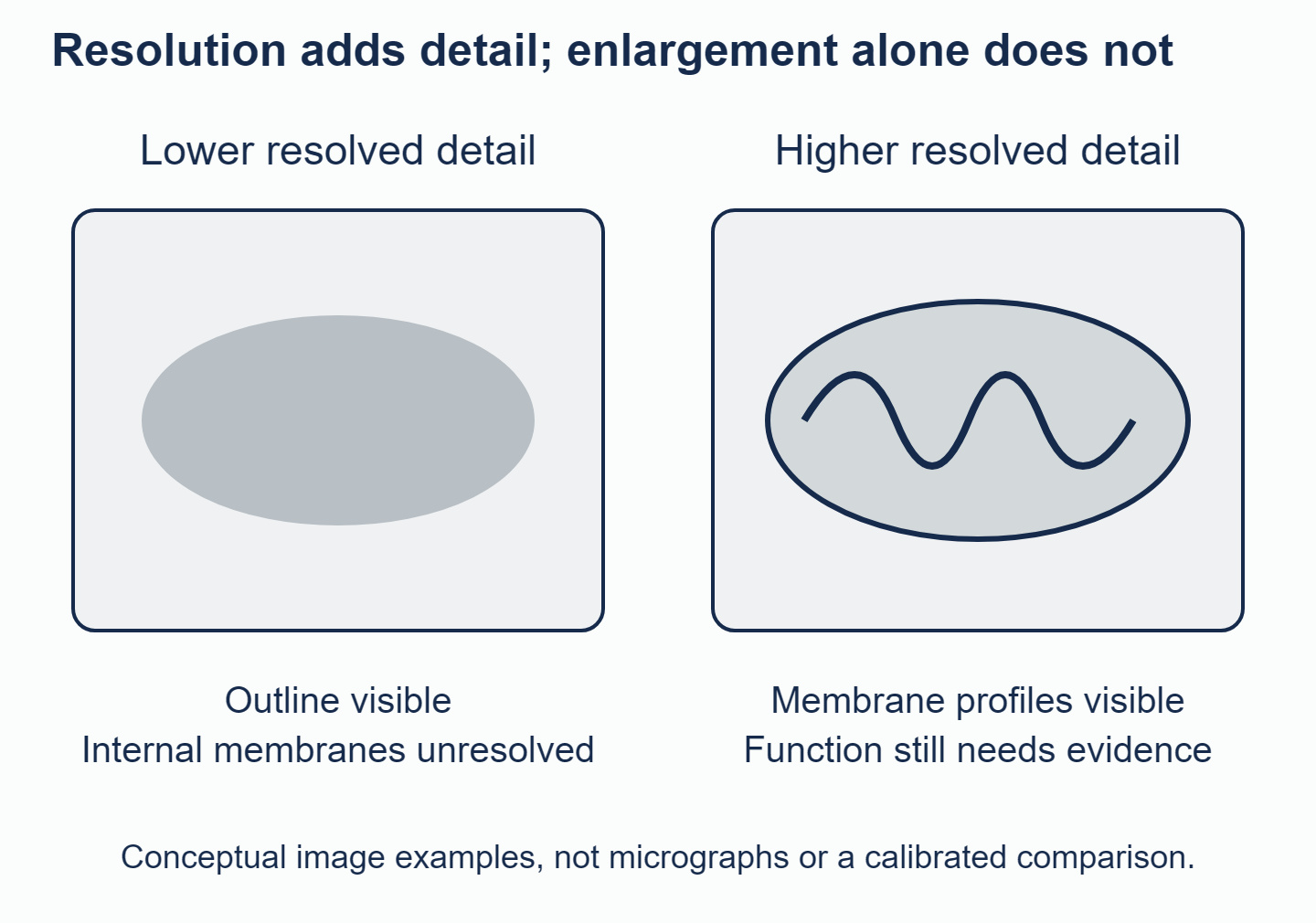
Seeing an outline differs from seeing internal detail; neither image directly measures how the structure works.
In transmission electron microscopy (TEM), electrons pass through a thin specimen. Different parts transmit different amounts, creating light and dark areas that reveal internal structures. For example, repeated membrane outlines inside a chloroplast help you identify its thylakoid system. A scanning electron microscope (SEM) typically scans a surface with a beam and detects signals that reveal its shape and texture. Specialised scanning methods can also image sections, so check the method rather than assuming every internal image is TEM.
The figure above is an explanatory schematic, not microscope evidence. With a real image, first check the method and scale bar, then distinguish what is visible from what you infer. A membrane fold is visible structure; the claim that it contains a particular enzyme usually needs additional biochemical or labelling evidence.
Even a sharp electron image has limits. Conventional preparation involves preserving the specimen (fixation), removing water, staining and cutting thin sections. Material can shrink, membranes can distort, and a knife mark can look like a biological boundary. These changes introduced during preparation are called artefacts. Cryogenic techniques, which use very low temperatures, reduce some problems but do not give an ordinary view of a living, functioning cell.
A thin section is also a two-dimensional sample. A tube cut across looks circular; another section through the same organelle may look elongated. An absent nucleus might simply lie outside the slice. Scientists can build a three-dimensional picture from successive slices, called serial sections, or from images taken at different angles using tomography.
Finally, shades of grey show differences in electron signals, not natural colour. A still image does not show which way or how fast a process happens. Live-cell light microscopy, fluorescent labels, repeated observations and experiments can help answer those questions. Use them alongside electron images to connect structure with function, while checking for preparation artefacts.
Explore the strengths and limits of electron images with the electron-microscopy question in the Biology Unity and Diversity questionbank. Support your evaluation with a structure that improved resolution can reveal and a question about cell function that a still image cannot answer on its own.
Answered by Revision Village IB Expert
Explore More IB Biology Resources
Over 80% of IB students globally are experiencing the power of Revision Village