Question
Biology
How do you calculate micrograph magnification from a scale bar?
Answer
Biology
Expert Answer
The magnification of a displayed or printed micrograph is the measured image length divided by the corresponding real length. A scale bar supplies a known real length, so it is usually the safest reference.
Both lengths must use the same unit. Magnification is a ratio, so the units cancel.
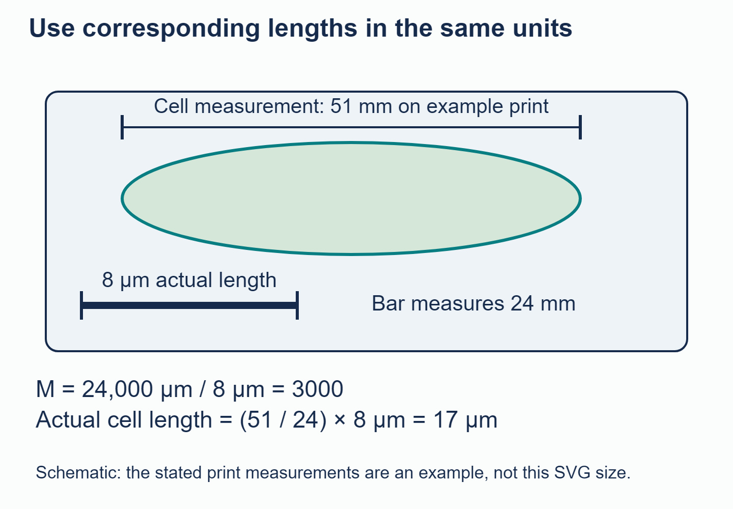
The cell-to-bar length ratio gives real size even when the complete image is resized.
Suppose a scale bar labelled measures on a particular printout. Convert millimetres to micrometres:
Therefore,
or . If a cell measures on that same printout, its real length is
You can reach the same answer directly from the length ratio:
This length-ratio method also works with pixels: if the cell spans 510 pixels and the scale bar spans 240 pixels, their ratio is unchanged. You do not need to convert pixels into millimetres to calculate the object's real size.
Resizing changes the image magnification. If the print is enlarged to twice its linear size, the bar becomes long and the image magnification becomes . The real cell remains because the cell and scale bar enlarge together. A copied caption saying only "" would now be misleading, whereas the embedded scale bar remains useful.
Do not confuse this with objective-lens magnification. A objective and eyepiece give a stated optical magnification of when you look through that microscope. Once an image is captured, its displayed magnification also depends on the camera sensor, display size, cropping and printing.
Measure between the actual ends of the bar, not between the ends of its text label. Keep the specimen and bar at the same zoom, and say which dimension you measured: the length along a cell's longest axis is different from its width. Finally, a larger image does not necessarily reveal more detail. That depends on resolution, the ability to distinguish close points as separate.
Build confidence with the microscopy question in the Biology Unity and Diversity questionbank. Set out the magnification method in your own words, making clear which lengths you measure and why their units must match before you divide.
Answered by Revision Village IB Expert
Explore More IB Biology Resources
Over 80% of IB students globally are experiencing the power of Revision Village