Question
Biology
How do you identify organelles in a plant-cell electron micrograph?
Answer
Biology
Expert Answer
Identify plant-cell organelles by combining clues from their location, boundaries and internal structure, rather than using size or darkness alone. An electron micrograph shows a section or surface of a prepared specimen. Some structures may therefore be outside the image or too unclear to distinguish.
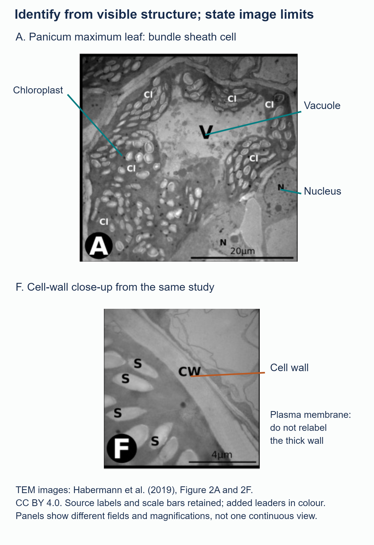
Panel A shows nucleus, vacuole and chloroplasts; panel F shows the cell wall. The panels have different scales, shown by the 20 μm and 4 μm scale bars.
Begin with the cell's outer boundary. The cell wall is a relatively thick layer outside the plasma membrane, separating neighbouring cell interiors. The plasma membrane is much thinner and lies just inside the wall. If you cannot distinguish a separate membrane line, identify the wall but explain that the membrane is not clear at this scale. Do not point to the thick wall and rename it "cell membrane".
A chloroplast contains an internal membrane system. Parallel membrane lines and stacks are stronger clues than an oval outline: mitochondria can also be oval. Chloroplasts may include starch granules, while mitochondria have folds of inner membrane called cristae, arranged differently. Also check what specimen you are looking at and the image's scale.
A vacuole is a compartment surrounded by a membrane and often looks relatively clear. A large clear region is a clue, but not enough on its own. Check that it lies inside the cell and has a suitable boundary; air spaces between cells are outside the cells, not vacuoles. The vacuole's membrane is called the tonoplast.
The nucleus is identified through its envelope and internal nuclear material. A nucleolus may appear as a denser region within it. A nucleus need not sit in the middle of a mature plant cell: a large vacuole can displace it towards the cell's edge. Nor must every section contain a nucleus.
Use a short evidence statement for each label: "chloroplast, because internal stacked membranes are visible" is stronger than "chloroplast, because it is dark". If a label is uncertain, say which feature is missing rather than inventing detail.
In panel A, the source labels N, V and Cl identify nucleus, vacuole and chloroplast respectively; the pale oval inclusions within chloroplasts are starch grains, not additional nuclei. Panel F supplies a more detailed wall view. These are different fields, with different scale bars, not a single continuous cell image.
Preparation changes dimensions and contrast; a static image establishes structure, not a photosynthetic rate.
Apply these clues to the tobacco leaf-cell micrograph in the Biology Unity and Diversity questionbank. For each labelled structure, explain which visible feature supports your identification and use it to rule out a tempting alternative.
<p class="image-attribution"><small>Image attribution: <a href="https://pmc.ncbi.nlm.nih.gov/articles/PMC6380572/" target="_blank" rel="nofollow noopener noreferrer">Habermann and colleagues (2019), Figure 2A and 2F</a>, reproduced under <a href="https://creativecommons.org/licenses/by/4.0/" target="_blank" rel="nofollow noopener noreferrer">CC BY 4.0</a>. Complete panels are selected, source annotations retained and new coloured labels added.</small></p>Answered by Revision Village IB Expert
Explore More IB Biology Resources
Over 80% of IB students globally are experiencing the power of Revision Village