Question
Biology
How do you identify starch, lipids and cellulose in a plant-cell micrograph?
Answer
Biology
Expert Answer
In a plant-cell micrograph, look for starch in storage granules within plastids, lipids in membranes and lipid-containing bodies, and cellulose in the cell wall. Plastids are organelles that include chloroplasts. Use the image to identify the likely structure first; its shade of grey alone usually cannot tell you which molecule it contains.
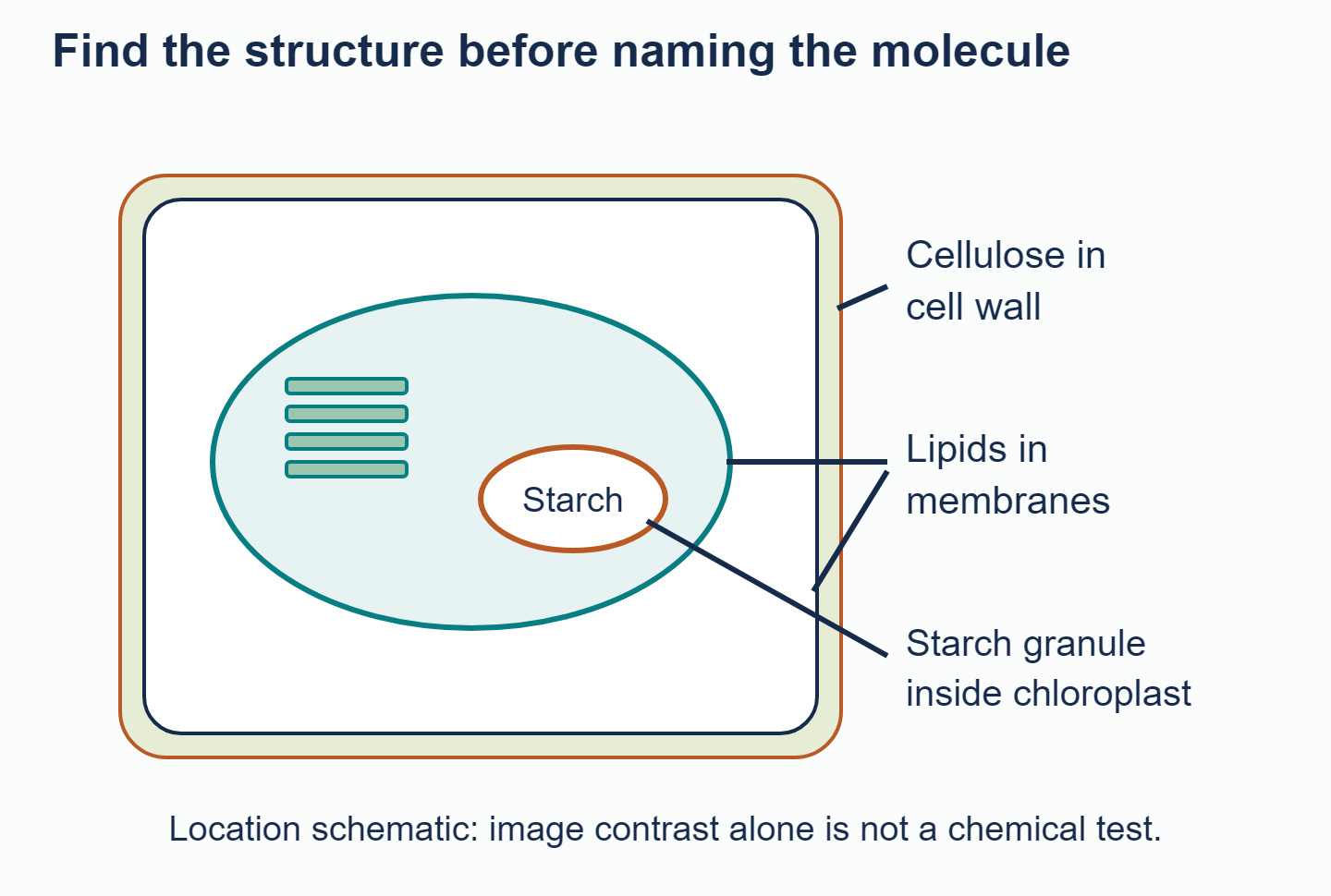
Starch granules, membranes and the cell wall are associated with different types of molecule.
Start by finding the chloroplasts. In a suitable electron micrograph, you can recognise their internal membranes, including stacks of thylakoids. Starch can collect in relatively large, pale granules in the stroma, the fluid-filled space around those membranes. In a photosynthetic leaf cell, a granule in the expected position with the expected appearance is reasonably identified as starch. A pale patch elsewhere could instead be a vacuole, an empty space or an artefact caused by preparing the specimen.
For lipids, a membrane is often a safer choice than an unidentified dark dot. The plasma membrane, chloroplast envelope, thylakoid membranes and vacuolar membrane all contain lipids. They contain different mixtures: photosynthetic membranes, for example, have many glycolipids, which are lipids with attached carbohydrate groups. Point your label to the visible membrane, not the empty-looking space it surrounds. A lipid droplet or a plastoglobule, a lipid-containing structure within a plastid, can also be suitable if the image supports that identification.
For cellulose, look outside the plasma membrane at the cell wall. Fine bundles of cellulose called microfibrils help strengthen the wall. It also contains other materials, so "cellulose is a component of the wall" is more accurate than "the wall is pure cellulose". The plasma membrane beside it has a different composition and function.
The diagram shows locations schematically; it is not an electron micrograph. When transferring the method to a real image, use each label's line endpoint carefully. A line ending in the chloroplast's starch inclusion differs from one ending in its envelope. A line crossing a wall but ending in the cytoplasm labels the endpoint, not everything along the line.
Finally, distinguish storage from structure. Starch consists of glucose polymers used for carbohydrate storage. Cellulose also consists of glucose units, but its different linkages permit strong structural fibres. Lipids contribute to membranes and can store energy. Identifying where a molecule belongs is a useful first step; confirming chemical identity may require specific staining or biochemical analysis.
Try locating these molecules in the Anemone leaf-cell micrograph in the Biology Unity and Diversity questionbank. Place each label carefully and give a structural reason for your choice, rather than relying only on whether an area looks light or dark.
Answered by Revision Village IB Expert
Explore More IB Biology Resources
Over 80% of IB students globally are experiencing the power of Revision Village